4.5 Scanning tunnelling microscopy
STM measurements were carried out at the School of Physics and Astronomy, University of Birmingham with assistance from Dr. Q. Guo and Mr. E. Green.
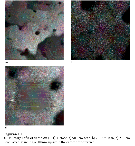
Gold was freshly evaporated onto mica and the quality of the surface verified by examination using the STM. The surface displayed the characteristic ‘herringbone’ reconstruction of Au (111) (figure 4.9).449 The step height is ~0.2 nm corresponding to a monatomic step. The dithiol porphyrin lacking b substituents, 230, was examined first. The gold substrate was immersed into a 0.4 mM solution of 230 in THF for 3 h, followed by rinsing with THF. The sample was imaged in air and revealed the surface to be covered with a ‘mat’ of material which could be displaced by scanning with the STM tip.
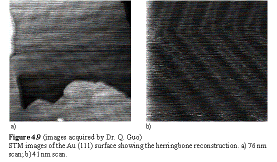
Figure 4.10 shows a series of scans in which the scan size was reduced then enlarged. It is clear that the tip has penetrated the layer on the surface and scraped out a 100 nm square. The apparent thickness of the layer, measured from a histogram of the height distribution of the image, is ~0.5 nm, although the real thickness of the surface layer will be greater than this due to the tip penetration. More extensive washing of the sample with THF, water and EtOH appeared to remove some of the loosely adsorbed material from the surface, although no ordered structures could be seen in the STM images. Irregular packing of 230 in a disordered layer on the surface may be responsible for the failure to observe structure in the STM images. Oxidative polymerization may also be a problem with this porphyrin leading to adsorption of a multilayer. The conclusion of a disordered layer or multilayer is in agreement with that reached for the b substituted analog 223 on the basis of the contact angle and electrochemical evidence.
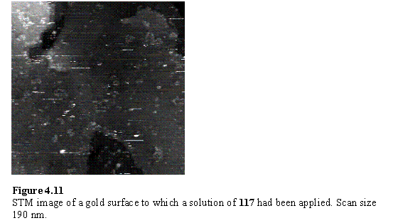
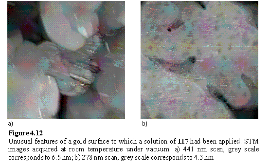
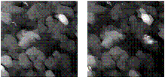
The literature reports examples of submolecular resolution STM images of aromatic compounds,450-452 such as porphyrins,453-459 physisorbed onto substrates such as graphite and metals. Hence it was decided to investigate STM imaging of porphyrin 117 directly on the Au(111) surface. The STM instrumentation was not capable of operation under a fluid environment so in situ imaging of 117 adsorbed from solution onto a surface was not feasible. However the facility to operate at elevated temperature under vacuum was available. It was hoped that application of a dilute solution of 117 to a gold surface, followed by drying and gentle heating might create a porphyrin layer which could be visualized by STM. A 10 mL drop of THF solution of 117 in which the red colour of the porphyrin was barely visible (concentration < 10-7 M) was applied to a newly prepared gold surface and the solvent allowed to evaporate in air. The surface was imaged under ambient conditions and revealed debris on the gold terraces (figure 4.11), although closer examination did not reveal any further structure that might indicate an ordered porphyrin array or otherwise. After application of a further 10 mL drop of porphyrin solution the system was placed under vacuum (~5 ´ 10-7 mbar) and the sample imaged again. Large parts of the surface had the appearance of bare gold terraces although some unusual striated features were observed (figure 4.12 a) along with terraces with a mottled appearance (figure 4.12 b).
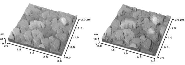
The temperature was progressively increased to 138 °C over a 7 h period, with regular imaging of the same area (figure 4.13). Some motion of the edges of the gold terraces was observed, and this is expected as it is known that thin films of gold on mica anneal at elevated temperature.460 A rough feature, similar to that of figure 4.12 a, was observed to grow during the course of the experiment. Possible origins could be outgassing or reaction of impurities buried under or within the film as the temperature was raised. Imaging of smaller areas of the terraces did not reveal any definitive features that could be ascribed with any certainty to porphyrin.
The failure to observe molecularly resolved images of porphyrins could be due to a combination of a number of factors. On drying of the THF solution of 117 the porphyrin may not form an even deposit on the surface but clump together in aggregates, such that most of the surface consists of bare gold. In support of this hypothesis AFM images (not shown) of THF solutions of 117 dried onto mica revealed flat featureless areas reminiscent of the bare mica surface and dome shaped structures of micrometre dimensions ascribed to the porphyrin. An alternative choice of solvent system may enable uniform films of porphyrin to be produced by evaporation. Another factor which may be responsible for the lack of molecularly resolved images may be the condition of the STM tip. The atomic configuration at the apex of the tunnelling tip determines the image quality, but this is largely outside the control of the experimenter. Adsorption, desorption and rearrangement of the atoms of the tip may change its condition during the course of scanning causing variation in the image quality. Successful imaging is likely to come with a combination of the correct tip and sample preparation, and a degree of luck.